
Image-guided Neurosurgery
fUS in the OR
Neurosurgeons continuously face life-altering decisions in an intra-operative setting. The imaging techniques that can guide them in making these decisions can be just as life-altering.
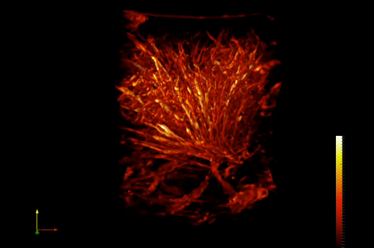
Oncological neurosurgery is an example of a speciality which relies heavily on making continuous, intra-operative tumor-brain delineations. Currently available imaging and mapping techniques such as (f)MRI, DTI and ESM present with serious limitations including lack of real-time intra-operative feedback, lack of depth resolution and risks of epileptic seizure elicitation. Functional ultrasound (fUS) presents as a new mobile neuro-imaging tool with unprecedented spatiotemporal resolution, which allows for possible identification of tumor borders based on vascular strucuture, as well as a distinction between healthy functional and unhealthy tumor tissue, based on differences in vascular response during particular functional tasks performed by the patient.