
Whole-brain Functional Imaging
Complete Exploration in High-Res
Unravelling the brain in all its complexity has been the goal of neuroscientists for centuries. Most of the tools available for high-resolution interrogation of the brain, force us to consider small regions of the brain at once, making it difficult to consider the bigger picture.
Motor planning, language processing, visual decoding, all functionalities of the brain we want to understand in health and cure in disease. Many if not all neuroscientific techniques we have available to measure brain activity in these phenomena, present with a compromise between the field of view, temporal and spatial specificity, sensitivity, and physical constraints on the animal. Functional Ultrasound (fUS) allows us to make whole-brain images of brain activity with unprecedented spatiotemporal resolution.
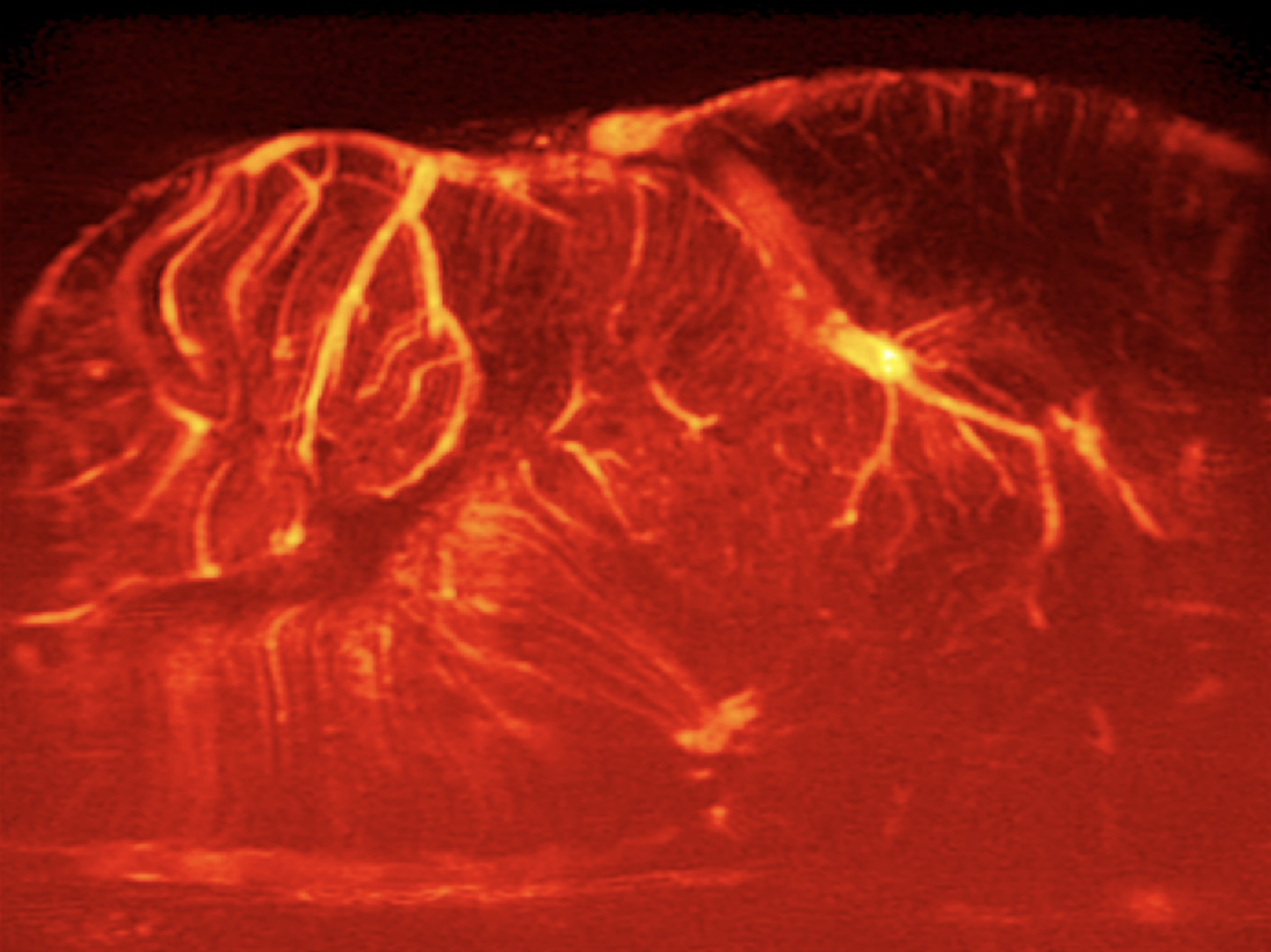
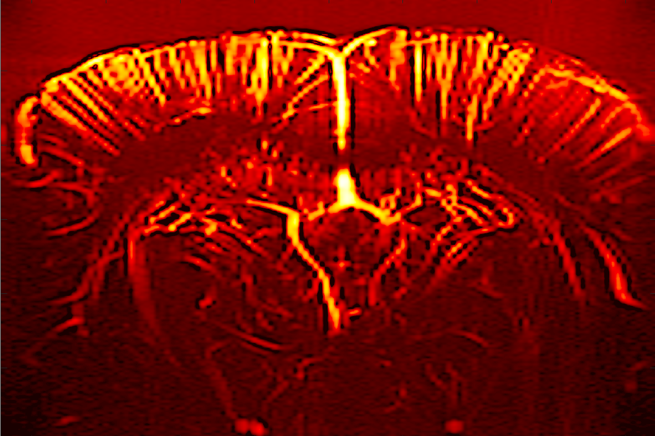
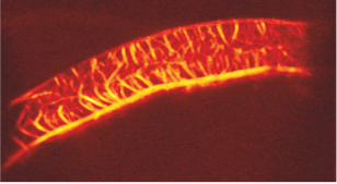
In fUS, detailed images of cerebral blood dynamics are captured by visualizing the minute motions induced by moving blood cells and possibly other vascular related motion such as dilation and vasoconstriction. Changes in the dynamics of the cerebral blood supply to specific locations may reflect local changes of metabolic activity upon neuronal activation. This process is referred to as neurovascular coupling (NVC). So far, fUS has been applied successfully in brains and spinal cords ranging from mice to humans, proving to be a powerful tool for studying the dynamics of endogenous brain signals.